Filter by Program Type:
Thursday, 12 Nov 2026
Speakers
-
 Dr Larissa Steigmann, Germany
-
 Dr Marius Steigmann, Germany
Abstract
Successful membrane coverage is determined by tension free flap closure and is significant for desirable clinical outcomes. Hence, proper tension release on the overlying flap to achieve passive tension flap closure remains to be the most important factor for achieving predictable bone augmentation outcomes. Flap openings have been associated with postsurgical complications including infections and graft failure. Therefore, gaining flap flexibility for the coverage of small to high volume augmentation is an important component for predictable outcomes. Several techniques have been described in the literature for flap advancement. Vertical releasing incision and periosteal scoring are common techniques to obtain flap flexibility and are commonly applied. Also, it is known that the incision into submucosa and even some muscularly incision will provide major flap advancement. Furthermore, the application of these advancement methods is often limited to the presence of sufficient gingival tissue thickness.
The determination of periodontal phenotype is relevant to determine mucogingival surgical management. For thin gingival tissue it has been advocated to perform a full thickness flap (periosteum within the flap) rather than a partial-thickness flap (leaving the periosteum over the bone as tissue thickness decreases wound dehiscence occurs. This is leading to flap tearing and subsequent graft exposure. Similar to flap advancement, split thickness flaps are surgical demanding to be performed in thin tissue as perforation may occur. So far, no specific surgical technique has been proposed for major flap advancement in the thin tissue phenotype (≤1.5mm). Therefore, this lecture introduces a surgical technique – The Mucosal Detachment Technique – that is separating the mucosal insertion from the periosteum which is aimed at achieving adequate dimensions of flap flexibility to cover high volume augmentation.
Learning objectives
- Explain the importance of tension-free flap closure for predictable membrane coverage and bone augmentation success.
- Identify causes and consequences of flap opening and graft exposure following augmentation procedures.
- Compare conventional flap advancement techniques and their limitations in thin periodontal phenotypes.
- Assess periodontal phenotype to guide appropriate mucogingival surgical management.
- Understand the principles and indications of the Mucosal Detachment Technique for achieving flap advancement in thin tissue cases.
Agenda
| 09:00 – 11:00 | Decision tree of soft tissue manipulation for site-specific GBR, differences in soft tissue manipulation between the anterior mandible, anterior maxilla, posterior mandible, and posterior maxilla - Dr Marius Steigmann |
| 11:00 – 11:30 | Coffee Break |
| 11:30 – 13:00 | Flap design and sutures in the non-aesthetic zone The mucosa detachment technique The periosteal pocket flap - Dr Larissa Steigmann |
| 13:00 – 14:00 | Lunch |
| 14:00 – 15:15 | Hands-on Site-specific soft tissue management for guided bone regeneration - Dr Larissa Steigmann & Dr Marius Steigmann |
| 15:15 – 15:30 | Coffee Break |
| 15:30 – 17:00 | Hands-on Different sutures technique and a complete GBR procedure. - Dr Marius Steigmann |
| 17:00 – 17:30 | Wrap-up & Q&A |
Supported by
No programs of this type on this day
Friday, 13 Nov 2026
Speakers
-
 Prof Ivan Darby, Australia
Abstract
Dental implants are now a routine part of modern dental practice. However, successful implant therapy extends far beyond implant placement alone. Following tooth extraction, significant dimensional changes of the alveolar ridge occur, particularly in the aesthetic zone, with ridge width reductions of up to 40%. Even with immediate implant placement, careful management of post-extraction remodelling is essential to preserve bone volume and achieve predictable functional and aesthetic outcomes.
Guided Bone Regeneration (GBR) is a well-established, evidence-based technique for reconstructing and preserving alveolar bone, providing the foundation for long-term implant success. This course explores the biological principles of GBR, clinical indications, surgical techniques, materials, and expected outcomes. During the hands-on workshop, participants will gain practical experience in GBR procedures for both delayed and immediate implant placement, including bone grafting and soft tissue management, to build confidence in applying these techniques in everyday clinical practice.
Learning Objectives
By the end of this course, participants will be able to:
- Explain the biological principles and clinical indications of Guided Bone Regeneration (GBR) in implant dentistry.
- Describe the healing processes of bone grafts and soft tissues following regenerative procedures.
- Identify the available grafting materials and barrier membranes, and discuss their clinical applications.
- Demonstrate the principles and techniques for the effective use of GBR materials during implant surgery.
- Demonstrate the clinical skills required for bone graft placement in immediate and delayed implant cases.
- Discuss soft tissue management techniques to optimise functional and aesthetic outcomes around dental implants.
Agenda
| 09:00 – 10:45 | Lectures Biological Principles of Bone Regeneration
|
| 10:45 – 11:15 | Coffee Break |
| 11:15 – 13:00 | Lectures GBR Around Implants
|
| 13:00 – 14:00 | Lunch |
| 14:00 – 16:45 | Hands On Participants will practise regenerative procedures around pre-positioned implant sites using simulated defects and soft tissue closure techniques.
|
| 16:45 – 17:00 | Discussion and Q & A |
Registration & PricingDownload BrochureWhatsapp Enquiry
Supported by

No programs of this type on this day
Saturday, 14 Nov 2026
Speakers
-
 Dr Giorgio Tabanella, Italy
Abstract
The management of the buccal bone remains one of the most critical challenges in implant site development. The Shield technique represents an evolution of the traditional socket preservation. In fact, the Shield technique is not only designed to preserve buccal contour while maintaining biological activity and vascular supply but also to properly augment the missing buccal bone at the time of tooth extraction. This approach has demonstrated how temporary, biologically active scaffolds can stabilize hard and soft tissues during early healing, minimizing postextraction collapse. However, anatomical variability, defect extension, and complex ridge deficiencies often require a more comprehensive regenerative strategy. Guided Bone Regeneration (GBR) has evolved accordingly, incorporating the same biological principles—space stability, angiogenesis, and controlled resorption—while expanding its indication range through advanced barrier technologies. In particular, resorbable magnesium membranes have introduced a new paradigm in GBR, bridging the gap between passive barrier function and active biological modulation. This presentation illustrates the conceptual and clinical transition from the Shield technique to modern GBR, framing both approaches as part of a single biological continuum rather than competing techniques. Clinical decision-making, material selection, and long-term tissue stability are discussed to support a rational, biology-driven regenerative workflow.
Learning Objectives
After attending this presentation, participants will be able to:
- Explain the biological principles underlying Shield technique using magnesium membranes.
- Recognize the advantages of magnesium-based barriers in buccal bone preservation and early healing.
- Identify clinical scenarios where the shield concept alone is insufficient and GBR is indicated.
- Understand GBR as an extension of the shield philosophy, rather than a separate or opposing approach.
- Implement a biologically coherent regenerative strategy based on defect morphology and tissue dynamics.
Speakers
-
 Dr Abdullah Al-Shamkhani, Iraq
Abstract
This lecture presents a practical framework for assessing dental implant cases by complexity and risk, using the ITI SAC classification (Straightforward, Advanced, Complex) to guide treatment planning. It examines the dental, medical, anatomical, and patient-related factors that shape case difficulty, alongside a structured risk-management approach: identifying, measuring, educating, mitigating, and reviewing potential complications. Through clinical case examples, the session demonstrates how thorough assessment and sound decision-making support safer, more predictable outcomes in implant dentistry.
Learning Objectives
- Discuss the principles of implant case assessment and classification
- Identify and evaluate patient-related, anatomical, medical, dental, and surgical risk factors that may influence implant treatment outcomes.
- Explain how clinician experience, treatment approach, biomaterial selection, documentation, and available equipment contribute to predictable implant therapy.
- Demonstrate a systematic approach to risk management through identification, assessment, patient education, risk mitigation, and clinical review.
- Analyze clinical implant cases to discuss treatment planning, management of complex situations, and strategies for achieving safer and more predictable surgical outcomes.
Speakers
-
Dr Giorgio Tabanella, Italy
This course is not available for individual purchase. If you wish to attend the course on 14 November 2026 (limited seats available only), you have to register for the conference (13-14 November) as the first one hour of the course is a lecture by Dr Giorgio Tabanella as part of the Clinical Implantology Symposium (CIS). The registration fee covers both days.
Abstract
The clinical success of regenerative procedures depends not only on biological principles but also on correct material handling and surgical execution. Magnesium membranes introduce a new regenerative paradigm by combining space maintenance with active biological interaction and controlled resorption, requiring a specific clinical protocol distinct from conventional collagen or nonresorbable barriers. This hands-on course is designed to provide participants with a step-by-step understanding of how to handle, shape, stabilize, and apply magnesium membranes in both the Shield technique and Guided Bone Regeneration procedures. Emphasis is placed on membrane positioning, fixation strategies, graft containment, and soft-tissue management to ensure predictable outcomes. Through guided practical exercises, participants will gain direct experience with magnesium membrane behaviour during hydration, adaptation to defect morphology, and integration within a regenerative workflow. The course aims to translate biological concepts into reproducible clinical actions, allowing clinicians to confidently incorporate magnesium membranes into daily practice.
Learning objectives
At the end of this hands-on course, participants will be able to:
- Understand the handling properties of magnesium membranes, including shaping and adaptation.
- Apply the Shield technique using a magnesium membrane for buccal bone preservation ed augmentation.
- Perform Guided Bone Regeneration procedures with magnesium membranes in 3D bony defects.
- Select appropriate fixation and stabilization techniques to ensure membrane stability during healing.
- Avoid common clinical errors and manage intraoperative challenges specific to magnesium membranes.
- Integrate magnesium membranes into a standardized clinical workflow for predictable regenerative outcomes.
Agenda
| 10:30 - 13:30 | Lecture: Clinical Application of Magnesium Membranes in Shield Technique and Guided Bone Regeneration |
| 13:30 - 14:30 | Lunch Break |
| 14:30 - 15:30 | Fundamentals and Shield Technique • Clinical orientation and biological principles • Indications and case selection • Magnesium membrane handling and shaping (demo + hands-on) • Shield technique workflow and execution (demo + hands-on) |
| 15:30 - 16:00 | Coffee Break |
| 16:00 - 17:00 | Guided Bone Regeneration and Clinical Application • GBR principles using magnesium membranes • Management of horizontal and vertical defects (demo + hands-on) • Fixation and stabilization concepts • Soft-tissue closure and troubleshooting • Key takeaways and Q&A |
Supported by
Speakers
-
 Dr Pokpong Amornvit, Thailand
Abstract
Implant dentistry has traditionally focused on bone availability, implant positioning, and osseointegration. However, achieving optimal outcomes requires a broader perspective that extends beyond the bone to include overall facial profile and esthetic harmony. A facial driven design approach emphasizes planning implant treatment based on facial references, ensuring that functional rehabilitation also contributes to improved facial proportions and appearance.
Rather than considering implant placement in isolation, clinicians must adopt a multidisciplinary strategy that integrates implantology, restorative dentistry, and orthodontics. This approach enables coordinated modification of tooth position, occlusion, and jaw relationships to achieve a more balanced and esthetically pleasing facial outcome. Implant placement may also serve as anchorage for clear aligner therapy, further enhancing treatment efficiency and predictability.
Advances in digital dentistry have made this integrated workflow more accessible and precise. Technologies such as intraoral scanners, facial scanners, and cone beam computed tomography (CBCT), combined with advanced planning software, enable clinicians to visualize and simulate treatment outcomes prior to surgery. These tools facilitate comprehensive treatment planning that aligns dental structures with facial esthetics.
This lecture presents the concept of facial driven design in digital implant dentistry, with a focus on pre-surgical planning to optimize facial outcomes. By combining clear aligner therapy, restorative procedures, and implant placement within a unified digital workflow, clinicians can achieve highly predictable, efficient, and esthetically driven results. The session highlights how digital tools support this multidisciplinary approach, ultimately improving both functional and facial outcomes for patients.
Learning Objectives
- Discuss the concept of facial driven design in implant dentistry and its clinical significance.
- Explain the importance of integrating facial analysis into implant treatment planning.
- Describe the multidisciplinary approach combining orthodontics, restorative dentistry, and implantology.
- Demonstrate how digital tools, including intraoral scanners, facial scanners, and CBCT, support facial driven workflows.
- Compare conventional implant planning with facial driven digital planning approaches.
- Apply facial driven design principles to enhance esthetic and functional outcomes in implant cases.
Speakers
-
 Dr Alessandro Venditti, Italy
Abstract
More and more colleagues are asking for solutions to restore edentulous patients with fixed teeth even in cases of severe atrophy and poor-quality bone. Flexibility, agility, competence. This is what it takes to master extreme cases.
This session is aimed at those who already face full-arch rehabilitations with confidence but often stop in front of the most challenging cases, especially in the atrophic maxilla. For those looking for solutions, this is the session for them. After this session, participants will increase their treatment options in cases of moderate and severe bone resorption, and will be able to treat more patients with an immediate full-arch protocol.
It will be shown how to solve full arch cases in one day, graftless and with immediate loading.
It will be discussed how to solve 100% of full arch cases with Maxilla-For-All® treatment concept by using pterygoid, trans sinus and zygomatic implants.
Learning Objectives
- Identify indications for graftless full-arch rehabilitation in atrophic maxillae.
- Explain the principles of the Maxilla-For-All® treatment concept.
- Differentiate the clinical applications of pterygoid, trans-sinus, and zygomatic implants.
- Demonstrate treatment planning for immediate full-arch loading in severe bone atrophy.
- Apply evidence-based protocols to manage complex full-arch cases in a single day.
Sponsored by:

Speakers
-
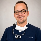 Dr Christoph Weinbach, Germany
Abstract
Immediate implant placement has become an increasingly important treatment approach in modern implant dentistry, particularly in the esthetic zone where preservation of hard and soft tissue architecture is critical for long-term success.
The presentation will discuss the biologic and aesthetic advantages of zirconia implants, including excellent soft tissue response, reduced plaque accumulation, biocompatibility, and their role in metal-free restorative concepts. Scientific evidence related to immediate implant placement, osseointegration, peri-implant tissue stability, and long-term esthetic performance will be reviewed with direct relevance to daily clinical practice.
Special attention will be given to the simplicity and efficiency of the SDS Swiss Dental Solutions implant system, emphasizing streamlined surgical and prosthodontic workflows that support minimally invasive treatment concepts and predictable restorative outcomes.
A major focus of the symposium will be the presentation of numerous clinical cases from the author’s daily practice, particularly in highly demanding anterior esthetic situations. Participants will gain practical insight into atraumatic extraction techniques, immediate implant placement protocols, soft tissue management, provisionalization concepts, prosthetic integration, and digital workflows designed to optimize esthetic and functional success.
Clinical recommendations for patient selection, treatment planning, risk assessment, and maintenance strategies will also be discussed to help clinicians improve predictability and long-term stability in immediate zirconia implant therapy.
By combining scientific evidence, digital innovation, and extensive clinical experience, this symposium aims to provide dentists, implantologists, prosthodontists, and restorative clinicians with practical tools and confidence to successfully implement immediate zirconia implant protocols in modern esthetic dentistry.
Learning objectives
- Participants will be able to discuss the biologic and esthetic advantages of zirconia implants in immediate implant placement protocols, particularly in the esthetic zone.
- Participants will gain the ability to explain the key principles of immediate implant placement, including atraumatic extraction, implant positioning, soft tissue preservation, and provisionalization strategies.
- Through clinical case presentations, participants will better understand and demonstrate the simplicity and efficiency of the implant system in both surgical and prosthodontic workflows.
- Participants will learn to evaluate treatment planning and risk assessment factors that influence predictable esthetic and functional outcomes with immediate zirconia implant therapy.
- By analyzing real-world clinical cases, participants will develop practical insight into digital workflows, prosthetic integration, and long-term maintenance strategies for immediate zirconia implant rehabilitations.
Speakers
-
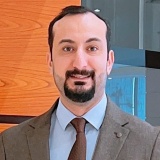 Dr Ahmed Aljumili, Iraq
Abstract
This lecture focuses on combining digital workflow with precise surgical execution in full-arch implant rehabilitation, with emphasis on implant positioning, soft tissue management, and achieving stable esthetic outcomes.
Learning Objectives
- Understand digital workflow in full-arch cases
- Improve implant positioning for better soft tissue outcomes
- Apply practical strategies for soft tissue management
- Enhance predictability in esthetic results
Speakers
-
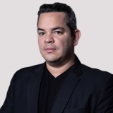 Dr Jesus Ostos, Spain
Abstract
Full-mouth implant rehabilitation demands a highly accurate and efficient digital workflow to achieve predictable functional and aesthetic outcomes. A comprehensive digital workflow has been developed to simplify and optimize every stage of the full-arch restorative process by integrating digital data acquisition, prosthetically driven planning, and seamless communication between the clinician and the dental laboratory.
This lecture will present a comprehensive digital workflow for full-arch implant rehabilitation, highlighting strategies to improve precision, reduce clinical and laboratory complications, and increase treatment efficiency.
Clinical cases will demonstrate how a fully digital approach enhances prosthetic accuracy, minimizes chairside adjustments, and improves passive fit, leading to more predictable long-term outcomes. Emphasis will also be placed on workflow standardization, interdisciplinary collaboration, and practical tips for integrating digital technologies into daily implant practice.
Learning Objectives
- Understand the principles of a fully digital workflow for full-mouth implant rehabilitation, from data acquisition to definitive prosthetic delivery.
- Apply digital protocols for accurate full-arch implant rehabilitation, including intraoral scanning and prosthetic-driven planning.
- Optimize communication between clinician and dental laboratory through digital integration to improve accuracy, efficiency, and predictability.
- Identify strategies to minimize clinical and prosthetic complications by improving implant position verification, passive fit, and restoration accuracy.
- Implement fully digital workflow into daily implant practice to standardize full-mouth rehabilitation and achieve more predictable long-term outcomes.
No programs of this type on this day
Sunday, 15 Nov 2026
Speakers
-
 Dr Marcus Dagnelid, Sweden
Abstract
This interactive workshop provides participants with a hands-on introduction to modern implant workflows using the latest technologies. Attendees will gain practical experience in optimizing implant placement through advanced motor control, torque management, and digital integration.
The session will cover key aspects of surgical workflow, including system setup, clinical protocols, and real-time decision-making during implant placement. Participants will explore how combining reliable implant systems with innovative surgical units can enhance precision, safety, and efficiency.
Through guided exercises and clinical demonstrations, clinicians will develop a deeper understanding of how to incorporate these technologies into daily practice, improving both surgical outcomes and workflow consistency.
Learning Objectives
- Explain the key components of modern implant workflows, including system setup, motor control, torque management, and digital integration.
- Discuss clinical protocols and real-time decision-making strategies essential for safe and predictable implant placement.
- Demonstrate proper implant placement techniques using advanced surgical units and implant systems during hands-on exercises.
- Explain how combining innovative surgical technologies with reliable implant systems enhances precision, safety, and clinical efficiency.
- Apply acquired skills to integrate modern implant workflows into daily practice, improving surgical outcomes and workflow consistency.
Agenda
| 09:00 – 10:30 | Introduction to Modern Implant Workflows Overview of current implant workflow concepts, including digital integration, motor control, and torque management. System Setup & Surgical Protocols Step-by-step walkthrough of equipment setup and clinical protocols for efficient and predictable implant placement. |
| 10:30 – 11:00 | Coffee Break |
| 11:00 – 12:30 | Hands-on Session I: Implant Placement Fundamentals Guided exercises focusing on:
|
| 12:30 – 13:30 | Lunch Break |
| 13:30 – 14:15 | Clinical Decision-Making Interactive session exploring intraoperative decision-making and strategies to enhance safety and outcomes. |
| 14:15 – 15:30 | Hands-on Session II: Integrated Workflow Practice Participants apply full workflow concepts, combining implant systems with surgical units to improve efficiency and consistency. |
| 15:30 – 15:45 | Coffee Break |
| 15:45 – 16:30 | Case Discussions Case-based discussions on optimising workflows in daily practice. |
| 16:30 – 17:00 | Q&A and Wrap-up Key takeaways, discussion, and guidance on implementing learned techniques into clinical routines. |
Supported by
No programs of this type on this day
Chairperson
Thank you to our chairperson for their support during the event.