Filter by Program Type:
Thursday, 12 Nov 2026
Speakers
-
 Dr Nur Saadeddin, Romania
-
 Dr Thomas Mendel, Romania
Abstract
The Er:YAG 2940 nm laser offers a versatile approach to a wide range of dental procedures involving both hard and soft tissues.
This lecture and hands-on workshop will provide an overview of the fundamental characteristics of the 2940 nm wavelength, laser–tissue interactions, ablation mechanisms, and essential safety considerations, with an emphasis on their clinical relevance.
Through clinical cases from the speakers’ daily practice, the multidisciplinary applications of the LIGHTMED Sapphire Er:YAG laser will be presented, including cavity preparation, zirconia crown debonding, gingivectomy and gingivoplasty, implant uncovering, periodontal debridement, biofilm disruption, granulation tissue removal, and surgical exposure of unerupted permanent teeth.
The hands-on session will focus on device handling, parameter and tip selection, and practical exercises demonstrating selected clinical procedures.
The program aims to highlight the versatility, ergonomics, and practical integration of Er:YAG laser technology into everyday dental practice.
Learning Objectives
Upon completion of this program, participantswill be able to:
- Understand the key characteristics of theEr:YAG 2940 nm wavelength and its interaction with dental tissues.
- Recognize the indications and clinicalapplications of Er:YAG lasers in different dental disciplines.
- Understand the principles of parameterselection, tip selection, and laser safety.
- Evaluate clinical techniques throughpractical case presentations.
- Gain hands-on experience with the LIGHTMEDSapphire Er:YAG laser system.
Agenda
| 11:30 – 13:30 | Part I – Er:YAG 2940 nm: Fundamentals and Clinical Principles
|
| 13:30 – 14:30 | Lunch Break |
| 14:30 – 16:00 | Part II – Multidisciplinary Clinical Applications Clinical cases and practical examples from the speakers’ daily practice:
|
| 16:00 – 16:30 | Break |
| 16:30 – 18:00 | Part III – Hands-On Workshop
|
Supported by
No programs of this type on this day
Friday, 13 Nov 2026
Speakers
-
 Dr Arun Darbar, UK
Abstract
Photobiomodulation (PBM) and Photodynamic Therapy (PDT) represent innovative, non-invasive treatment modalities increasingly utilised in modern dentistry. PBM employs low-level light therapy to stimulate cellular activity, enhance tissue repair, and reduce inflammation, proving beneficial in procedures such as pain management, wound healing, and the treatment of oral mucositis. Conversely, PDT involves the application of a photosensitizing agent activated by a specific wavelength of light, producing reactive oxygen species that effectively target and eliminate pathogenic microorganisms.
This abstract explores the synergistic potential of PBM and PDT within dental practice. The review outlines their mechanisms of action, clinical applications, and the therapeutic outcomes observed in periodontal therapy, endodontic treatments, and oral lesion management. Furthermore, it discusses the advantages of these therapies, including reduced antibiotic reliance, minimal side effects, and improved patient comfort. Despite the promising benefits, the abstract also acknowledges existing challenges, such as standardization of protocols and the need for further clinical trials to substantiate efficacy.
In conclusion, the integration of PBM and PDT into dental protocols signifies a progressive shift towards more biologically driven, patient-centred care strategies, with substantial implications for improving oral health outcomes.
In our practice, we have now implemented the next feature of PBM and PDT with the same wavelengths, incorporating modified clinical protocols in clinics. This advancement has been complemented by motivated patient uptake, supported through continued home care units and structured maintenance phases. Additionally, early clinical observations indicate improved patient outcomes and adherence, highlighting the potential for broader application in diverse therapeutic settings. Continuous monitoring and feedback mechanisms have been integrated to refine efficacy, ensuring sustained patient engagement and optimized treatment results.
Learning Objectives
- Gain a thorough understanding of the primary principles and theories relevant to the subject matter.
- Develop the ability to implement learned concepts in real-world scenarios effectively.
- Cultivate analytical skills to evaluate information critically and make informed decisions.
- Strengthen both verbal and written communication to articulate ideas clearly and confidently.
- Foster teamwork abilities to work efficiently within diverse groups and contribute meaningfully.
- Encourage adaptability and creative thinking to address challenges and embrace new approaches.
- Promote continuous self-evaluation to identify personal strengths and areas for improvements.
Speakers
-
 Dr Imneet Madan, UAE
Abstract
The Calm Chair Method is a behavioral framework designed to change how children experience dental care. Children are not typically scared of the dentist itself — they fear not being heard and understood. By facilitating practice with modalities that replace fear with emotional empowerment, motivation, and strength-building, dental appointments can become serene memories.
The Calm Chair Method combines elements of NLP (Neuro-Linguistic Programming), Early Years Foundation Stage principles, child development psychology, Buteyko breathing, EFT (Emotional Freedom Technique), Theta Healing, Psych-K®, and laser dentistry. Once a child is emotionally settled, a laser-assisted, needle-free approach can then be used. Lasers as the capstone not only transform fear, but also help maintain trust and confidence — with lasers as the capstone, calm becomes the first treatment, and connection the first cure.
Speakers
-
 Dr Fadi Khuffash, UAE
Abstract
Ankyloglossia, commonly known as tongue-tie, is a congenital condition characterized by a restrictive lingual frenulum that limits tongue mobility. Despite its high prevalence, estimated between 0.1% and 10.7% of the population, tongue-tie remains underdiagnosed and its far-reaching consequences on pediatric development are often overlooked. This lecture aims to reshape the clinician's understanding of ankyloglossia by exploring its multidimensional impact beyond the commonly recognized breastfeeding difficulties.
The lingual frenulum plays a fundamental role in stabilizing the tongue and facilitating essential oral functions including breathing, swallowing, speech, and proper tongue posture. When restricted, the tongue cannot exert the physiological forces necessary for normal palatal expansion and maxillofacial growth. Emerging evidence demonstrates that untreated tongue-tie contributes to maxillary constriction, narrow palatal vaults, mandibular prognathism, Class III malocclusion, mouth breathing, and compromised airway development in growing children.
This presentation provides a comprehensive overview of the etiology and classification of ankyloglossia, followed by an in-depth analysis of its consequences on craniofacial development, speech articulation, and overall pediatric growth. Furthermore, the lecture critically compares two primary frenectomy approaches: conventional surgical release using a scalpel, which requires suturing and heals by primary intention, versus laser-assisted frenectomy utilizing CO2, Nd:YAG, or diode lasers, which achieves simultaneous hemostasis, eliminates the need for sutures, and heals by secondary intention with reduced postoperative pain and faster recovery.
By integrating current scientific evidence with clinical applications, this lecture will equip practitioners with the knowledge to identify tongue-tie early, understand its developmental implications, and select the most appropriate release technique based on patient age, clinical severity, and long-term treatment goals. This is not merely a lecture about a minor procedure — it is a paradigm shift in how we perceive the tethered tongue.
Learning Objectives
- Explain the anatomy, embryology, and physiological function of the lingual frenulum and its critical role in tongue posture, palatal development, and craniofacial growth.
- Identify the etiology and risk factors of ankyloglossia, including congenital, genetic, and developmental origins.
- Describe the impact of tongue-tie on pediatric craniofacial development, including maxillary constriction, mandibular prognathism, malocclusion, narrow palate, and impaired jaw growth.
- Recognize the clinical indications for frenectomy, encompassing breastfeeding difficulties, speech impediments, airway obstruction, mouth breathing, and orthodontic considerations.
- Compare laser and conventional surgical frenectomy techniques, evaluating differences in bleeding control, pain management, operative time, and suturing requirements (primary vs. secondary intention healing).
Speakers
-
 Dr Fernando Duarte, Portugal
Abstract
Among the different causes of implant failure — most of which relate to clinical aspects of the implant treatment, medications, and health conditions — bacterial periodontal infections have been reported to be a major risk factor for early and late loss of dental implants. Post-extraction periodontal pathogens have also been linked to the etiology of peri-implantitis and periodontitis, and to increased bone loss, reduced bone healing, and implant instability.
Current methods for post-extraction disinfection of the implant site are mainly based on drugs and surgical techniques, although the pharmacological risk of the former and the invasiveness of the latter have led researchers to seek alternative, safer therapeutic methods — one of which is laser therapy.
This lecture outlines new laser protocols consisting of degranulation, disinfection, decortication, de-epithelialization, clot stabilization, and photobiomodulation using Er:YAG and Nd:YAG wavelengths to prevent and manage peri-implantitis clinical cases.
Speakers
-
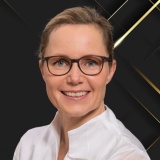 Dr Gabriele Schindler-Hultzsch, Germany
Abstract
Modern pediatric dentistry increasingly demands an interdisciplinary approach to efficiently manage complex orofacial anomalies, developmental disorders and traumatic dental injuries, always considering the special demands of the pediatric patient. The combination of innovative technologies such as Erbium- and Diode lasers, cone-beam computed tomography (CBCT), microsurgical techniques, early orthodontic intervention, and AI-assisted diagnostics opens new possibilities for diagnosis and treatment planning in pediatric patients. The aim of this work is to illustrate the potential benefits of an advanced multidisciplinary treatment concept in pediatric dentistry through selected clinical cases and to highlight the synergies between the individual modalities. A case series of 12 pediatric cases with different indications was retrospectively analysed, involving CBCT-guided three-dimensional diagnosis of impacted, dislocated teeth or supernumerary teeth followed by laser-assisted surgical exposure with Er,Cr:YSGG (2780nm) and Diode Laser (940nm), orthodontic alignment with AI-based automated growth prediction from digital radiographs, and interdisciplinary management of trauma patients involving combined surgical-orthodontic therapy supported by laser-assisted wound and pulp tissue treatment, with follow-up ranging from 4 months to 3 years. In all cases, the combination of technologies resulted in more precise diagnostics, reduced treatment times and improved patient compliance. CBCT enabled exact three-dimensional localization of anatomical structures, while AI algorithms significantly supported diagnostic accuracy and the predictability of orthodontic growth patterns. Laser therapy markedly reduced intraoperative and postoperative discomfort. The multidisciplinary approach incorporating modern technologies represents a promising paradigm shift in pediatric dentistry. The integration of AI, imaging, surgery, and orthodontics fosters personalized, efficient, and child-friendly treatment. Further prospective studies with larger case numbers are necessary to confirm the long-term clinical relevance and cost-effectiveness of this integrative concept.
Learning Objectives
- Discuss the advantage of CBCT for the approach and procedure of minimally invasive laser-assisted surgery.
- Name three different therapy options of an impacted tooth.
- Discuss the treatment options available in cases of trauma with a root fracture.
- Explain the advantages of a multidisciplinary approach for complex cases in pediatric dentistry.
- Identify which laser wavelengths can be used for surgical procedures and the advantages of Erbium wavelengths.
Speakers
-
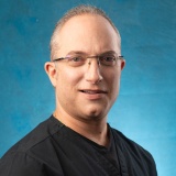 Dr Isaac Kably Mizrahi, Mexico
Abstract
Laser-assisted endodontics has evolved from a technology primarily used for disinfection into a biologically driven therapeutic approach capable of influencing the root canal environment, periapical tissues, and the host healing response. Understanding the biological mechanisms underlying laser–tissue interactions is essential for optimizing clinical outcomes while ensuring safety and predictable treatment.
This lecture will review the current evidence regarding the biological effects of different laser wavelengths used in endodontics, including their interaction with dentin, irrigants, microorganisms, and periapical tissues. Particular emphasis will be placed on laser-enhanced irrigation dynamics, biofilm disruption, smear layer modification, dentinal tubule decontamination, and the preservation of surrounding tissues. The presentation will also explore emerging evidence on photobiomodulation, inflammatory modulation, macrophage polarization, angiogenesis, mitochondrial activation, and their potential contribution to periapical healing and postoperative comfort.
The lecture will critically examine the scientific literature supporting laser-assisted endodontic procedures, highlighting both established evidence and areas where further research is needed. Clinical protocols, treatment parameters, and safety considerations will be discussed to facilitate evidence-based integration of laser technologies into contemporary endodontic practice.
By combining biological principles with clinical applications, participants will gain a comprehensive understanding of how laser-assisted endodontics may enhance treatment outcomes beyond conventional chemomechanical preparation while recognizing the limitations of current evidence.
Learning Objectives
- Explain the biological principles of laser–tissue interactions relevant to endodontic therapy.
- Discuss the effects of different laser wavelengths on dentin, irrigants, biofilms, and periapical tissues.
- Describe the biological mechanisms involved in laser-enhanced disinfection, smear layer modification, and dentinal tubule decontamination.
- Evaluate the current scientific evidence supporting laser-assisted endodontics, distinguishing established findings from emerging concepts.
- Demonstrate how biological principles can be translated into evidence-based clinical protocols for laser-assisted endodontic treatment.
- Discuss the potential role of photobiomodulation in modulating inflammation, promoting tissue repair, and improving postoperative outcomes.
- Identify key safety considerations and clinical parameters required for the predictable and responsible use of lasers in endodontics.
Speakers
-
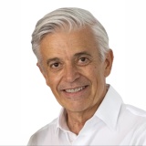 Dr Kresi Simunovic, Switzerland
Abstract
The presentation focuses on a non-invasive laser assisted anti-snoring therapy, the evolution and 15 years of clinical experience, the protocols, outcomes, challenges and limitations.
It includes the basic knowledge requirements in sleep medicine as dentist for a better communication with professionals and patients, and a possible first detection of apneas and hypopneas in the dental office.
The main focus is the treatment of primary snoring and eventual support in case of obstructive sleep apneas and hypopneas, the goal is improvement and not always absolute silence, explained by some patient cases.
Learning Objectives
- Discuss the dentist's role in the early detection of sleep disorders and sleep apnea.
- Explain the principles and protocol of laser-assisted anti-snoring therapy.
- Describe the fundamentals of sleep medicine for effective interdisciplinary communication.
- Differentiate primary snoring from obstructive sleep apnea and hypopnea.
- Identify the goals, indications, and limitations of laser-assisted anti-snoring therapy.
Speakers
-
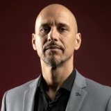 Dr Luciano Mayer, Brazil
Abstract
Dental laser therapy has traditionally been structured around single-wavelength protocols. Although highly effective for specific indications, this approach may not fully address the biological complexity encountered when multiple tissues, therapeutic objectives, and healing mechanisms coexist within the same clinical procedure. Recent advances in laser technology now allow the simultaneous delivery of multiple wavelengths, creating new opportunities for biologically integrated treatment strategies. This lecture introduces a biologically driven clinical framework for simultaneous multi-wavelength laser treatment, demonstrating how different wavelengths can be strategically integrated according to tissue characteristics, biological targets, and clinical objectives. The framework is developed from current scientific evidence, principles of laser-tissue interaction, and extensive clinical experience, integrating photothermal, photomechanical, and photobiomodulatory effects into a coherent decision-making process — emphasizing tissue-specific responses, parameter selection, procedural sequencing, and safety considerations across surgical, periodontal, implant, and rehabilitative procedures. The proposed framework demonstrates how simultaneous multi-wavelength application allows complementary biological effects to be integrated within a single clinical workflow. Rather than selecting wavelengths independently, treatment planning becomes centered on biological targets, tissue behavior, and desired therapeutic responses, contributing to greater procedural consistency, improved biological modulation, and enhanced clinical predictability. The future of laser dentistry is not defined by the availability of additional wavelengths, but by a deeper understanding of biological targets and laser-tissue interactions. Simultaneous multi-wavelength treatment provides clinicians with a biologically integrated strategy that refines clinical reasoning and supports more predictable, evidence-based, and patient-centered care.
Learning Objectives
- Explain the biological principles supporting simultaneous multi-wavelength laser treatment.
- Interpret laser-tissue interactions according to biological targets rather than isolated wavelengths.
- Design biologically driven multi-wavelength treatment strategies based on tissue characteristics and therapeutic objectives.
- Integrate surgical laser applications and photobiomodulation within a single clinical workflow.
- Critically evaluate the current scientific evidence supporting simultaneous multi-wavelength approaches in contemporary dental practice.
Speakers
-
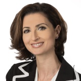 Dr Marina Polonsky, Canada
Abstract
In this presentation, we will discuss the different wavelength options available and how to choose the appropriate device to achieve the desired treatment goals and additional services dentists may want to add to their clinical practice. We will review the principles of laser–tissue interactions and identify which lasers are best suited for efficient cutting, coagulation, disinfection, soft-tissue surgery, and hard-tissue applications. Lasers are not all created equal. When clinicians understand the intended clinical outcome and the physical principles that determine how lasers behave in living tissues, we can consistently deliver an improved patient experience. Soft tissue surgical and non-surgical procedures, minimally invasive treatment of periodontal pockets, photo-bio-modulation in the supportive treatment of dentinal hypersensitivity and the management of TMJD are easy procedures for beginners and utilize entry level diode lasers. Hard tissue applications like laser-activated irrigation of complex root canal systems, laser cavity preparation with reduced need for local anesthesia are possible with other lasers. Understanding how lasers can improve clinical outcomes and the patient experience will inform, engage, and inspire attendees to incorporate lasers into their clinical practice.
Learning objectives
- Learn about different types of lasers used in dentistry and their applications.
- Explain important considerations when purchasing your first laser.
- Discuss laser–tissue interactions important for hemostasis vs ablation.
- Learn what makes lasers an irreplaceable instrument in the dentist’s armamentarium.
- Demonstrate how patient experience can be enhanced when lasers are used instead of conventional tools
Speakers
-
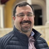 Dr Masoud Mojahedi, Iran
Abstract
The rapid emergence of antibiotic-resistant bacteria is a global challenge, threatening the effectiveness of antibiotics that have transformed medicine and saved millions of lives.
While the treatment of carious lesions and their consequences was once the primary focus of dental care, periodontal disease and its associated complications are now considered a leading cause of tooth and bone loss in many patients.
This presentation aims to highlight the advantages of laser-assisted periodontal therapy and its role in improving infection control and treatment outcomes.
Laser light technologies for infection control can help reduce the need for antibiotics while improving clinical outcomes in periodontal therapy.
Learning Objectives
- Identify the different laser types and parameters used to achieve antibacterial effects in dentistry.
- Describe the clinical applications and techniques for using lasers in dental practice.
- Evaluate the role of lasers as an adjunct to conventional dental treatment.
- Discuss the antibacterial efficacy of lasers in the management of oral and dental conditions.
- Recognize the limitations, safety considerations, and potential thermal effects associated with laser use in dentistry.
Speakers
-
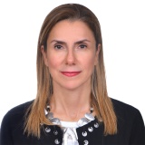 Prof Ruya Yazicioglu, Türkiye
Abstract
The integration of laser technology into restorative dentistry has expanded significantly, offering innovative and minimally invasive approaches for the management of hard dental tissues. Laser systems are increasingly utilized for cavity preparation, selective ablation of caries, and surface modification to enhance restorative material adhesion. Laser irradiation can effectively remove the smear layer, create microretentive surface topographies, and induce physicochemical alterations in dental substrates, thereby influencing bonding performance and potentially reducing the need for conventional mechanical instrumentation.
Beyond hard tissue applications, lasers have also demonstrated clinical value in the management of dentin hypersensitivity through mechanisms such as dentinal tubule occlusion, protein coagulation, and modulation of neural responses. Furthermore, in aesthetic dentistry, laser-assisted tooth bleaching has gained attention for its ability to accelerate the activation of bleaching agents, shorten chair-side treatment time, and improve patient comfort.
In summary, laser technologies in restorative dentistry offer more accurate tissue management and enhanced patient comfort, which supports minimally invasive concepts. Lasers can be a useful complement to conventional methods when utilized with the right indications and parameters. However, careful case selection, operator expertise, and adherence to evidence-based protocols are necessary for long-term therapeutic success.
Learning Objectives
- Discuss the current clinical applications of lasers in hard tissue procedures and their advantages and limitations
- Describe the mechanisms and clinical indications for laser-assisted management of dentin hypersensitivity
- Evaluate the effectiveness, advantages, and limitations of laser-assisted tooth whitening compared with conventional bleaching techniques
- Identify appropriate indications and treatment parameters for incorporating lasers into restorative dental practice
- Recognize the safety considerations and clinical parameters required for the predictable and effective use of lasers in restorative dentistry
Speakers
-
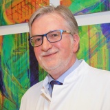 Prof Siegfried Jänicke, Germany
Abstract
The application of lasers in medicine and dentistry is based on the interaction between biological tissues and laser light radiation. Shortly after Maiman and colleagues (USA) developed the first functional solid-state laser in 1960, the initial medical applications of lasers were explored.
The use of lasers in dentistry can also be traced back to 1965. As frequently the introduction of novel therapeutic procedures in medicine, the new methodology initially saw a comparatively widespread, and at times uncritical, application. Only gradually did fields such as ophthalmology, dermatology, vascular malformations, and other surgical applications emerge as primary indications for lasers within medicine.
In dentistry, the focus was predominantly on the potential of lasers for painless cavity preparation, eliminating the unpleasant side effects of traditional ‘drilling’. However, due to device sizes and costs, such therapeutic approaches were initially largely restricted to scientific research. It was not until approximately 1989 that the adoption of lasers in American and German dental practices gradually commenced.
In addition to laser therapy itself, diagnostic laser applications in medicine and dentistry must also be taken into account. Furthermore, other laser applications, such as the processing of biomaterials, are gaining increasing importance. This presentation aims to show the current focal points and intersections of laser applications in medicine and dentistry.
A look beyond national borders reveals that the US Food and Drug Administration (FDA) addresses the use of lasers in medicine and dentistry in detail on its website. It is striking that among the four main areas of laser application in medicine, dental therapies—such as endodontic or periodontal procedures, teeth whitening, or oral surgery indications—are mentioned as a matter of course. This is noteworthy because a quasi-official listing of dental laser therapy alongside other medical laser applications does not exist in Germany.
Learning Objectives
- Discuss the evolution of laser applications in medicine and dentistry.
- Explain the principles of laser–tissue interaction in clinical practice.
- Identify current therapeutic and diagnostic applications of dental lasers.
- Describe the evidence-based indications for laser use across dental specialties.
- Discuss current trends and future directions in laser dentistry.
Speakers
-
 Dr Ana Cecilia Aranha, Brazil
Abstract
This comprehensive lecture offers attendees a meticulous exploration of the applications of laser dentistry, providing insights into the multifaceted clinical applications of advanced laser technologies. Through case studies, participants will gain insights into the transformative potential of utilizing high-power diode lasers in various dental procedures.
The versatility of this technology offers a spectrum of benefits, including precise tissue incision with reduced post-operative discomfort and accelerated healing times. Emphasizing the advantages of targeted energy delivery and minimal thermal damage, attendees will discover how integrating high power diode lasers into clinical practice enhances treatment outcomes while optimizing patient comfort and satisfaction. Through an immersive examination of authentic clinical scenarios, participants will gain invaluable scientific insights, enabling them to seamlessly integrate laser dentistry and photobiomodulation into their clinical practices with precision and efficacy.
Learning Objectives
- Explain the mechanisms of laser-tissue interaction.
- Discuss the clinical applications of lasers in dentistry.
- Identify appropriate clinical indications for laser use.
- Demonstrate the selection of appropriate laser settings and application techniques.
- Apply evidence-based principles to support clinical decision-making in laser dentistry.
Speakers
-
 Dr Manoela D. Martins, Brazil
Speakers
-
 Dr Wolfgang Manschiebel, Austria
Abstract
Disinfection of the implant surface remains one of the greatest challenges in the non-surgical treatment of peri-implantitis. Limited access to the implant surface compromises the cleaning efficacy of various decontamination techniques, and even under controlled laboratory conditions, available in vitro studies generally fail to achieve complete biofilm removal. This may, at least in part, explain why clinically relevant differences between various mechanical and/or chemical decontamination methods remain difficult to demonstrate in clinical studies.
Shock Wave Enhanced Emission Photoacoustic Streaming (SWEEPS®) is an innovative technique for cleaning and disinfecting the root canal system in endodontics. Using a patented technology, an optimized sequence of shock waves is delivered into an irrigating solution, thereby enhancing the penetration of the irrigant and enabling its antimicrobial and biofilm-removing effects to reach lateral canals and microscopic dentinal tubules.
Given the success of SWEEPS® in endodontics, the micro-rough surface characteristics of dental implants, and the persistent difficulty of efficiently removing biofilm from these surfaces, the SWEEPS® technology represents a promising approach for implant surface decontamination in the non-surgical treatment of peri-implantitis. This concept will be investigated in the present in vitro laboratory study.
Learning Objectives
- Discuss why complete biofilm removal from the implant surface is a prerequisite for successful non-surgical peri-implantitis therapy.
- Describe how photoacoustic laser therapy (SWEEPS®) achieves complete removal of biofilm from micro-rough implant surfaces.
- Review the findings of the in vitro study and the clinical outcomes associated with this photoacoustic approach.
Speakers
-
 Dr Amir Ezzat, Egypt
Abstract
Implant dentistry has evolved beyond achieving osseointegration to optimizing every stage of treatment for improved biological outcomes, patient comfort, and long-term success. Laser technology has become an important adjunct throughout the implant workflow, offering minimally invasive solutions that enhance surgical precision, accelerate healing, and support the management of peri-implant tissues. This lecture provides a practical, evidence-based overview of laser applications before, during, and after implant placement. Topics include laser-assisted decontamination of extraction sockets, management of infected sites, photobiomodulation, soft tissue surgery, second-stage implant uncovering, and soft tissue conditioning during the restorative phase to optimize functional and esthetic outcomes. Particular emphasis will be placed on the prevention and management of peri-implant diseases. Beyond reviewing treatment protocols, the lecture will address a common clinical challenge: When should peri-implantitis be treated with an Er:YAG laser, and when should it not? Evidence-based criteria for case selection—including defect configuration, implant surface characteristics, extent of bone loss, and patient-related factors—will be discussed to help clinicians identify cases that are most likely to benefit from laser therapy and recognize situations where alternative surgical or regenerative approaches are more appropriate. Current scientific evidence will be combined with practical treatment algorithms and clinical cases to illustrate indications, limitations, laser parameters, and common pitfalls. By the end of this session, participants will have practical protocols and clinical decision-making tools that can be readily incorporated into daily implant practice, ultimately improving treatment predictability, peri-implant tissue health, and the long-term success of dental implants.
Learning Objectives
- Discuss how lasers can be integrated into every stage of implant therapy — from treatment planning to long-term maintenance
- Explain the different clinical uses of lasers before, during, and after implant placement to improve healing, patient comfort, and treatment outcomes
- Recognize when an Er:YAG laser is the right choice for treating peri-implantitis — and when other treatment approaches are more appropriate
- Demonstrate a practical approach to selecting laser protocols based on clinical findings, implant conditions, and patient factors
- Apply simple clinical protocols and treatment strategies that can be incorporated into everyday implant practice to improve the long-term health and success of dental implants
Speakers
-
 Dr Ana Catarina Nogueira da Silva, Portugal
Abstract
Successful adhesive dentistry depends on creating a durable bond between restorative materials and dental hard tissues. This lecture explores how laser irradiation parameters—including energy, fluence, pulse duration, frequency, and irradiation technique—affect tissue interaction and influence adhesive performance. Through current scientific evidence and clinical experience, participants will gain practical insights into optimizing bond strength, hybrid layer quality, nanoleakage, and adhesive system selection for laser-treated dentin. The session concludes with clinically applicable recommendations to improve restoration longevity, minimize adhesive failures, and support predictable, minimally invasive restorative outcomes.
Learning Objectives
- Understand how laser irradiation parameters influence dental tissue and adhesive performance.
- Evaluate the impact of laser settings on bond strength, hybrid layer quality, and nanoleakage.
- Select appropriate adhesive systems for laser-treated dentin based on current evidence.
- Apply practical laser and adhesive strategies to improve restoration longevity and clinical success.
- Integrate minimally invasive, evidence-based laser techniques into daily restorative practice.
Speakers
-
 Dr Anna Maria Yiannikos, Cyprus
Abstract
For decades, advances in laser dentistry have focused primarily on the optimization of physical parameters, including wavelength, fluence, pulse duration and tissue interaction. While these developments have significantly improved clinical outcomes, considerable inter-individual variability in healing and treatment response remains, even when identical laser protocols are applied.
This lecture proposes a complementary perspective: that the next major advance in laser dentistry may not come from further refinement of laser technology alone, but from a deeper understanding of the biological environment into which laser energy is delivered.
Drawing upon emerging evidence from oral microbiome research and host inflammatory biology, the presentation explores the distinction between microbial taxonomy and microbial function, highlighting that understanding what microorganisms are doing may become as important as knowing which are present.
Current evidence indicating that laser-based interventions may modulate biological healing processes, including inflammatory signaling, angiogenesis and tissue repair, will be critically reviewed, together with the limitations and heterogeneity of the existing literature.
Building upon these observations, the lecture introduces the concept of Biology-Guided Laser Dentistry as a proposed research framework for future investigation rather than a validated clinical model, in which treatment strategies may integrate conventional clinical assessment with biologically meaningful, patient-specific characteristics such as microbial ecology, functional biology and host response.
Rather than presenting definitive answers, the lecture aims to stimulate a new research dialogue on how biological intelligence may shape the future evolution of laser dentistry.
Learning Objectives
- Explain why optimization of physical laser parameters alone does not fully account for the inter-individual variability observed in healing and treatment response.
- Distinguish between microbial taxonomy ("which microorganisms are present") and microbial function ("what microorganisms are doing") in the context of oral health.
- Critically evaluate the current evidence suggesting that laser-based interventions may modulate biological healing processes, while recognizing the heterogeneity and limitations of the existing literature.
- Discuss the potential contribution of the oral microbiome, functional biology, host inflammatory response and the oral-systemic interface to future laser research.
- Appraise the concept of Biology-Guided Laser Dentistry as a proposed research framework rather than a validated clinical model.
- Identify future research directions through which biologically informed approaches may contribute to more personalized laser dentistry.
Speakers
-
 Dr Julia Kozlova, Russia
Abstract
Until nowadays the problem of finding the most effective method for root canals instrumentation and chemical irrigation is still open. Researchers are trying to find the easiest and simplest way to perform endodontic treatment and most importantly it should be effective in long term follow up.
There are different techniques to use lasers during endodontic treatment. One concept is to activate irrigation solution (Sodium hypochlorite or EDTA) – it is called laser-activated irrigation (LAI). Another technique is to use distillate water as an irrigation solution and 2 wavelengths: combination of Erbium and diode lasers. Erbium lasers are used for removing the smear layer to open dentin tubules and diode lasers in dry root canal for anti-microbiological effect.
Erbium lasers effectively remove the smear layer during working regarding caries and non-carious lesions treatment and before final obturation in endodontic treatment.
In our research from 2019 Er:YAG showed high anti-microbiological potential during endodontic treatment. Research showed effectiveness of Er:YAG lasers in removing smear layers during endodontic treatment. Due to laser selectivity and targeting only chromophores depending on the wavelengths of laser used we could speak about minimal invasive dentistry as a one page of modern biological dentistry.
Learning Objectives
- Discuss dentin surface modification in contemporary adhesive protocols.
- Explain the advantages of laser-assisted caries preparation.
- Describe the double-wave concept and its clinical applications.
- Demonstrate laser applications from endodontics to restorative dentistry and compare them with conventional techniques.
- Apply laser-assisted techniques to achieve minimally invasive and conservative treatment outcomes.
Speakers
-
 Dr Cristina Neculau, Romania
Abstract
Advanced peri-implantitis associated with extensive circumferential bone loss and Grade III implant mobility is generally considered to have a poor prognosis, with explantation often regarded as the most predictable treatment option. Whether peri-implant bone support can be biologically re-established through a strictly non-surgical laser protocol, without grafting materials, remains insufficiently documented.
Learning Objectives
- Describe the clinical characteristics and CBCT findings of advanced peri-implantitis associated with extensive bone loss and Grade III implant mobility.
- Explain the biological rationale for combining Er,Cr:YSGG laser decontamination with 940 nm photobiomodulation in a non-surgical treatment approach.
- Discuss the clinical protocol used for laser-assisted management of a severely compromised implant without grafting materials or membranes.
- Interpret the clinical and CBCT findings that documented peri-implant bone re-establishment and long-term implant stability during the 24-month follow-up.
- Evaluate the potential indications, limitations and clinical implications of biologically driven laser-assisted therapy in the management of advanced peri-implantitis.
Speakers
-
 Dr Koushik Naik, UK
Abstract
Immediate implant placement can reduce treatment time and support preservation of the existing alveolar architecture. However, maintaining interdental papillae and peri-implant soft tissue stability remains an important challenge, particularly in the aesthetic zone. Atraumatic extraction, effective socket debridement and management of the early healing environment are therefore important considerations in achieving favourable aesthetic and biological outcomes.
This clinical case series presents three cases of immediate implant placement incorporating a dual-wavelength laser protocol aimed at supporting extraction socket debridement and disinfection, soft tissue preservation and post-operative healing.
Following atraumatic tooth extraction, the sockets were debrided and disinfected using a 2940 nm Er:YAG laser at 3 W. Laser-assisted debridement was used to remove residual granulation tissue and decontaminate the extraction socket while minimising mechanical trauma to the surrounding hard and soft tissues. Immediate implant placement was subsequently performed with particular attention to preservation of the existing gingival architecture and interdental papillae.
Following implant placement, photobiomodulation (PBM) was performed using a 650 nm laser at 100 mW for 40 seconds. The PBM protocol consisted of three treatment sessions at three-day intervals, with the aim of supporting early tissue healing and modulating the post-operative inflammatory response.
Clinical and photographic follow-up across the three cases showed favourable healing with maintenance of the interdental papillae and peri-implant soft tissue architecture.
This case series illustrates the potential role of combining Er:YAG laser-assisted socket debridement and disinfection with post-operative photobiomodulation as adjuncts to a tissue-preserving immediate implant protocol. Further controlled clinical studies with standardised soft tissue measurements are required to evaluate their contribution to long-term peri-implant tissue stability and aesthetic outcomes.
Learning Objectives
- Discuss the biological and clinical factors influencing interdental papilla preservation and peri-implant soft tissue stability following immediate implant placement.
- Explain the principles and clinical rationale for using a 2940 nm Er:YAG laser for extraction socket debridement and disinfection.
- Demonstrate a tissue-preserving clinical workflow integrating atraumatic extraction, laser-assisted socket management and immediate implant placement.
- Describe the biological principles of photobiomodulation and its potential role in modulating inflammation and supporting early peri-implant soft tissue healing.
- Explain the clinical application of a 650 nm photobiomodulation protocol following immediate implant placement, including treatment parameters and timing.
- Evaluate the clinical soft tissue and interdental papilla outcomes demonstrated across three immediate implant cases.
- Discuss the potential benefits, limitations and current evidence considerations when integrating Er:YAG socket treatment and photobiomodulation into immediate implant protocols.
Speakers
-
 Dr Pilar Martin, Spain
Abstract
Oral soft-tissue restrictions in newborns and infants, including ankyloglossia and other lesions, may interfere with effective breastfeeding, tongue mobility, swallowing, and the physiological development of the orofacial structures. Early diagnosis and appropriate treatment are therefore essential to restore function and support normal orofacial development. Laser technology has emerged as a valuable tool for the management of these conditions due to its precision and favorable postoperative course.
To evaluate the clinical application, predictability, and postoperative outcomes of laser-assisted soft-tissue surgery in newborns and infants presenting with oral soft-tissue restrictions that may compromise breastfeeding or normal orofacial function.
A comprehensive pediatric oral examination was performed before treatment to assess tongue mobility, oral anatomy, functional restrictions, and the suitability of each patient for surgical intervention. Different laser wavelengths were selected according to the clinical indication, including diode lasers of 450 nm, 940 nm, and 980 nm, and an Er,Cr laser of 2780 nm. Clinical protocols included appropriate infant positioning and stabilization techniques, protective eyewear adapted for infants and clinical staff, topical anesthesia when indicated, and the use of hyaluronic acid gel as part of postoperative care.
In our clinical experience of more than 300 treated cases, laser-assisted soft-tissue surgery demonstrated a high degree of clinical predictability and effective intraoperative management. The procedures allowed precise tissue management with favorable postoperative healing and good clinical tolerance. The use of laser technology facilitated the management of delicate oral structures in very young patients and contributed to a controlled postoperative period. Clinical improvement in tongue mobility and breastfeeding function was observed in appropriately selected patients.
Laser-assisted treatment of oral soft-tissue restrictions can be a valuable clinical tool in newborns and infants. When combined with comprehensive diagnosis and appropriate patient selection, laser surgery may facilitate the restoration of functional tongue mobility, support the initiation or maintenance of breastfeeding, and contribute to the prevention of functional disturbances that could negatively influence normal orofacial development.
Learning objectives
- Explain the main oral soft-tissue restrictions that may affect breastfeeding and orofacial function in newborns and infants.
- Discuss the clinical criteria for diagnosis and appropriate patient selection for laser-assisted soft-tissue surgery.
- Describe the clinical principles and protocols involved in laser treatment of oral soft tissues in very young patients.
- Demonstrate appropriate clinical considerations for infant positioning, protection, anesthesia, and postoperative care.
- Evaluate the clinical outcomes, advantages, and limitations of laser-assisted treatment in newborns and infants.
Speakers
-
 Dr Saleh Aria, UK
Abstract
Dental implant therapy has become a predictable treatment modality; however, achieving optimal precision during surgery, maximizing primary stability, and promoting rapid soft- and hard-tissue healing remain key determinants of long-term success. The integration of dental laser technology into implantology has introduced new opportunities to enhance surgical accuracy, minimize tissue trauma, and improve postoperative outcomes.
This lecture will explore the clinical role of laser-assisted implantology across multiple stages of implant treatment, including soft-tissue management, bone preparation, implant site decontamination, and peri-implant tissue modulation. Particular emphasis will be placed on the use of all-tissue laser systems, such as the Er,Cr:YSGG 2780 nm wavelength, which enables precise interaction with both soft and hard tissues while preserving surrounding biological structures.
The presentation will discuss how laser energy can facilitate minimally invasive surgical approaches, enhance visibility through improved haemostasis, reduce bacterial contamination, and stimulate photobiomodulation effects that support faster healing and improved patient comfort. Clinical cases will demonstrate the application of lasers in immediate implant placement, implant site preparation, peri-implantitis management, and soft-tissue contouring.
Evidence-based principles, laser-tissue interaction mechanisms, and practical clinical protocols will be reviewed to help clinicians integrate laser technology safely and effectively into implant treatment workflows. By combining advanced laser technology with modern implant concepts, clinicians can achieve greater surgical precision, improved implant stability, and enhanced biological healing responses.
Learning Objectives
- Understand the principles of laser-tissue interaction relevant to implant surgery
- Identify the clinical advantages of laser-assisted implantology in both soft- and hard-tissue procedures
- Apply laser protocols for implant site preparation, soft-tissue management, and peri-implant decontamination
Speakers
-
 Dr Manaf Taher Agha, UAE
Abstract
Although dental lasers are increasingly available in clinical practice, many clinicians remain uncertain about their appropriate indications and practical value. Educational material often focuses on device physics and capabilities rather than real clinical decision-making. This lecture aims to provide a balanced, evidence-guided perspective on the everyday use of lasers in dentistry.
Through a series of representative clinical cases, the presentation will illustrate situations in which laser application can enhance treatment efficiency, optimize soft-tissue management, and improve patient comfort. Clinical examples will include soft-tissue surgery, laser-assisted restorative procedures, adjunctive periodontal therapy, implant-related interventions including peri-implantitis management, laser-assisted endodontic procedures, veneer debonding, tooth bleaching, and photobiomodulation for temporomandibular disorders and symptomatic oral mucosal lesions. Conventional treatment approaches will be discussed alongside each application to clarify relative advantages, limitations, and realistic clinical expectations.
Particular emphasis will be placed on matching wavelength selection to tissue characteristics and treatment objectives, while also identifying scenarios where laser use offers minimal clinical benefit. The goal is to provide clinicians with a practical framework for integrating laser technology into daily practice in a predictable and responsible manner.
Learning Objectives
- Identify common clinical situations in which dental lasers can improve treatment efficiency, soft-tissue management, and patient comfort in everyday practice.
- Differentiate the appropriate clinical indications for laser use in procedures including soft-tissue surgery, periodontal therapy, implant-related treatments, restorative procedures, endodontic applications, and photobiomodulation.
- Select appropriate laser wavelengths and treatment approaches based on tissue characteristics and clinical objectives.
- Recognize the limitations of laser use and determine when conventional treatment methods remain the preferred option.
- Integrate laser technology into routine dental workflows in a predictable and clinically responsible manner.
Speakers
-
 Dr Pilar Blanco, Colombia
Abstract
Surgical procedures involving soft tissues inevitably require tissue incision, resulting in tissue injury that activates physiological mechanisms responsible for wound repair and regeneration. Current evidence suggests that the biological response of the treated tissue is directly influenced by the cutting instrument used. For this reason, laser devices have been increasingly adopted in dentistry for more than five decades as an alternative to the conventional scalpel, aiming to overcome its limitations by providing improved hemostasis, reduced postoperative discomfort, and enhanced surgical precision. Despite their extensive clinical use, the biological effects of dental lasers have been investigated predominantly in animal models, while evidence derived from human oral tissues remains limited. Consequently, there is a need to characterize the histological effects of different laser wavelengths on human oral mucosa and correlate these findings with clinical outcomes. Such information is essential to better understand the tissue response induced by laser-assisted surgery and to identify the advantages and potential limitations of these technologies in periodontal practice. The present study aims to evaluate the cellular and microstructural changes in gingival tissues following gingivectomy performed with a 450 nm diode laser and a 2,940 nm Er laser, and to compare the histological and clinical outcomes associated with each wavelength. The findings are expected to provide clinically relevant evidence to support the selection of the most appropriate laser system for soft tissue surgery in dentistry.
Learning objectives
- To describe the sociodemographic characteristics of the study population.
- To identify the cellular and microstructural changes in the epithelium and connective tissue of the gingival biopsy specimens.
- To compare the histological findings in gingival biopsies obtained following gingivectomy performed with two different laser wavelengths: a 450 nm diode laser and a 2,940 nm Er:YAG laser.
- To describe the clinical outcomes associated with gingivectomy performed using the two different laser wavelengths.
- To determine whether significant differences exist in the histological characteristics of gingival tissues treated with a 450 nm diode laser versus a 2,940 nm Er:YAG laser.
Speakers
-
 Assoc Prof Youssef Sedky, Egypt
Abstract
Laser technology is increasingly expanding the boundaries of modern orthodontics, offering clinically relevant applications beyond conventional treatment protocols. This presentation will explore the emerging role of lasers in orthodontics, with particular emphasis on photobiomodulation therapy (PBMT) using diode lasers for treatment acceleration and pain control.
A major focus will be the importance of appropriate laser parameters—including wavelength, power, energy density, exposure time, and treatment protocol—as key determinants of biological response and clinical success. The presentation will progress from the biological principles and current evidence supporting PBMT to practical, clinically applicable protocols, illustrated through clinical cases and evidence-based findings.
The second part will introduce a less explored application of erbium laser technology in orthodontics, focusing on its potential role in the rebonding of ceramic brackets. Clinical cases will demonstrate the application of erbium lasers in managing bracket debonding and rebonding, while selected clinical and laboratory studies will provide insight into its effectiveness, safety, and potential advantages over conventional techniques.
By connecting scientific evidence with real-world clinical applications, this lecture aims to provide orthodontists with a practical understanding of how laser technology can be integrated into contemporary orthodontic treatment, while highlighting emerging opportunities for more efficient, comfortable, and biologically driven care.
Learning objectives
- Explain the biological principles of photobiomodulation in orthodontics.
- Discuss the role of diode lasers in accelerating orthodontic treatment.
- Identify appropriate laser parameters for pain control and clinical effectiveness.
- Describe clinical protocols for integrating PBMT into orthodontic practice.
- Evaluate the use of erbium lasers for ceramic bracket debonding and rebonding.
Speakers
-
 Prof Dr Walid Altayeb, Qatar
Abstract
Excessive gingival display “gummy smile” seems to be one of the main esthetic feature of the periodontium that plays an important role on oral health-related quality of life and can have an adverse effect on the patient’s perception of attractiveness and self-confidence. Altered passive eruption APE is one of the causal factors of Excessive gingival display. Esthetic crown lengthening surgery could be considered a predictable protocol for APE and achieving more harmonious and symmetrical smiles.
Esthetic crown lengthening surgery to manage APE could include gingivectomy and/or apically repositioned full-thickness flap, with/without ostectomy. The width of keratinized gingiva, the position of the gingival margins, the location of the buccal alveolar crest, the location of the mucogingival junction, and the likelihood of concomitant restorative therapy are all factors that collectively determine the esthetic crown lengthening approach. Clinical application of lasers in periodontal surgery has continued to expand in the last decade and the concept of minimally invasive dentistry can be achieved by choosing laser treatment. The ability of erbium lasers to ablate both hard and soft tissues with excellent surgical precision and minimal collateral effects creates the opportunity for a minimally invasive flap and flapless, resulting in decreased tissue damage, minimize the bleeding, causing less inflammation, less post-operative discomfort, and may prevents collateral tissue damage that could occur with conventional methods.
Laser-assisted flapless esthetic crown lengthening may be a more patient-friendly and efficient procedure compared to traditional open-flap surgery with similar esthetic results. This could be important for dentists looking to offer their patients a less invasive and potentially more comfortable option.
Learning Objectives
- Understanding of the classification and treatment planning for gummy smile.
- Explain the biologic laser tissue interaction in periodontal tissues
- Discover how to meet the esthetic parameters through crown lengthening.
- Demonstrate the surgical techniques when performing esthetic crown lengthening with lasers
- Compare the clinical outcomes of erbium laser-assisted open-flap and flapless esthetic crown lengthening.
Speakers
-
 Dr Miguel Rodrigues Martins, Portugal
Abstract
Several laser wavelengths have been proposed and used in endodontics to enhance clinical outcomes and overcome the limitations of conventional irrigation protocols.
This lecture will present the scientific background and clinical approach to the use of the 445 nm laser in a variety of complex endodontic scenarios.
Discover how this simple and effective laser-assisted endodontic technique can transform and enhance your daily clinical practice.
Learning Objectives
- Explain the biophysical principles of laser wavelengths in endodontics.
- Discuss the clinical advantages of the 445 nm blue laser in endodontics.
- Describe the significance of deep laser penetration in endodontic treatment.
- Demonstrate the clinical applications of the blue laser in endodontic procedures.
- Discuss the management of iatrogenic complications using laser-assisted techniques.
Speakers
-
 Prof Marcia Martins Marques, Brazil
Abstract
This keynote highlights the contributions of our research group in Brazil to advancing the scientific foundations of photobiomodulation (PBM) in regenerative medicine. Over the past two decades, our work has bridged fundamental laboratory discoveries with preclinical and clinical applications, helping establish PBM as a scientifically grounded regenerative therapy.
Our research has demonstrated that PBM extends beyond inflammation modulation and analgesia by actively regulating cellular behaviour. We have shown that PBM promotes proliferation, migration, survival, extracellular matrix synthesis, and lineage-specific differentiation, providing a strong biological basis for tissue regeneration.
A central focus of our investigations has been mesenchymal stem cells (MSCs). Our studies revealed that appropriately dosed PBM enhances MSC proliferation, viability, metabolic activity, osteogenic differentiation, and mineralised matrix formation without inducing phototoxicity. We also demonstrated the importance of the biphasic dose response, showing that wavelength, energy density, power, and irradiation time are critical determinants of therapeutic outcomes and require careful optimisation for reproducible clinical protocols.
Building on these discoveries, we integrated PBM with tissue engineering strategies, including biomaterial scaffolds, bone substitutes, and stem cell-based therapies. These studies demonstrated that PBM enhances cell attachment, proliferation, differentiation, angiogenic signalling, and extracellular matrix deposition, significantly improving the regenerative performance of engineered tissues.
Our translational research provided compelling evidence supporting PBM in bone regeneration, demonstrating enhanced osteoblast maturation, collagen synthesis, osteogenic gene expression, and mineralized tissue formation. These findings were validated in animal models and translated into clinical applications in implant dentistry, guided bone regeneration, periodontal regeneration, oral surgery, and wound healing.
Beyond our scientific discoveries, we contributed to the 2025 World Association for Laser Therapy (WALT) Position Paper on PBM and Stem Cells, providing evidence-based recommendations and priorities for future clinical research. Collectively, our work has helped position PBM as an evidence-based therapeutic platform with significant potential to advance regenerative medicine.
Learning Objectives
- Discuss the biological mechanisms of photobiomodulation in tissue regeneration.
- Explain the effects of photobiomodulation on mesenchymal stem cell function.
- Describe the influence of PBM parameters on therapeutic outcomes.
- Demonstrate the clinical applications of PBM in regenerative dentistry.
- Discuss current evidence supporting PBM in tissue engineering and regenerative medicine.
Speakers
-
 James D. Carroll, UK
Abstract
Global life expectancy has increased by approximately 30 years over the past century, but gains in health span have not kept pace. Many individuals now spend the final 10–20 years of life living with chronic disease, disability, and declining functional capacity. Strategies that simultaneously target multiple age-related biological pathways may offer opportunities to improve both longevity and health span. This lecture reviews the mechanistic rationale and clinical evidence supporting photobiomodulation (PBM) as a potential intervention for improving physiological factors associated with healthy ageing, longevity, and age-related disease risk.
A narrative review was conducted of published clinical studies investigating the effects of PBM on physiological markers associated with cardiovascular health, metabolic function, physical performance, recovery, autonomic regulation, sleep quality, and inflammation, drawing on randomised controlled trials and higher-quality non-randomised clinical studies involving local and whole-body PBM interventions.
Across multiple studies, PBM demonstrated beneficial effects on several recognised predictors of health span and longevity, including improvements in exercise capacity, muscular strength, physical recovery, sleep quality, and quality of life. PBM has also been associated with reductions in systemic inflammatory and oxidative stress markers, improvements in heart rate variability and resting heart rate, reductions in nocturnal hypertension, and improvements in VO₂max, a well-established predictor of all-cause mortality.
PBM should not be viewed as a treatment for ageing itself, nor as a panacea for age-related disease. However, current evidence suggests that PBM may represent a useful adjunctive intervention capable of targeting several physiological domains associated with healthy ageing. Further large-scale prospective studies are warranted to determine whether improvements in recognised surrogate markers translate into meaningful long-term reductions in morbidity, disability, and mortality.
Speakers
-
 Dr Ilay Maden, UK
Abstract
Effective biofilm removal is fundamental to periodontal and implant therapy. Deep periodontal pockets, intrabony defects, extraction sockets, and complex implant surfaces can be challenging to clean thoroughly using mechanical methods alone.
To present a novel Er:YAG laser-induced photoacoustic approach for non-contact biofilm disruption and cleansing in periodontal and implant therapy.
Photoacoustic energy generates dynamic fluid movement and cavitation capable of disrupting biofilm without direct laser contact with the treated surface. Clinical applications include non-surgical periodontal therapy, surgical cleansing of intrabony defects, extraction socket management, cleansing of implant sites before implant placement, and treatment of peri-implantitis. The approach is designed to access complex anatomical areas while preserving surrounding soft tissues, root surfaces, bone, and implant surfaces.
Photoacoustic biofilm removal provides a versatile, non-contact adjunct for periodontal and implant site cleansing. Its ability to disrupt biofilm in difficult-to-access areas while supporting a tissue-preserving approach may expand the role of laser-assisted therapy in contemporary periodontal and implant care.
Learning objectives
- Explain the principles of Er:YAG laser-induced photoacoustic biofilm disruption in periodontal and implant therapy.
- Discuss the role of non-contact photoacoustic cleansing in managing biofilm within complex periodontal and peri-implant environments.
- Identify clinical indications for photoacoustic cleansing in non-surgical and surgical periodontal therapy.
- Demonstrate the clinical application of this approach in intrabony defects, extraction sockets, and implant sites prior to implant placement.
- Describe its application in the management of peri-implantitis and biofilm-contaminated implant surfaces.
Speakers
-
 Dr Grace Sun, USA
Abstract
More than half all Americans experience pain. 20-30% of these people suffer from orofacial pain. As health care practitioners we need to manage the pain as well as addressing the etiology in order to bring about the best outcome for the patient.
While current, traditional treatment modalities are both respected and important, an integrative approach is essential, including PBMT, Acupuncture and Traditional Chinese Medicine (TCM), chiropractic medicine, osteopathic medicine etc.
PBM, or PhotoBioModulation treatment, a therapeutic form of red and infrared light therapy, has come to be a unifying term covering low level laser treatment (LLLT), sourced from laser and/or LED light devices. PBM therapy increases healing efficiency, including reduction of inflammation and faster muscle recovery. Acupuncture, a complimentary pain management modality, stimulates neurological responses including endorphin release and by activating the meridian channel, reversing stagnation (pain) and increasing the flow of vital energy. These two modalities are in fact interdependent, complimenting and fortifying one another’s benefits. The meridian system can complimentarily be activated with PBM, when directed to the correct location. Meridian mapping techniques, the identification of correlated meridian points and anatomicalmapping techniques, the identification of correlated meridian points and anatomical landmarks, will be discussed.
Learning Objectives
- Understand the significance of exploring alternative approaches to pain management
- Learn integrative treatment approaches, including PBM therapy and acupuncture
- Review the functional mechanisms of PhotoBioModulation and Acupuncture
- Learn how to integrate PBM into your patients’ home care program
- Apply evidence-based principles to develop integrative PBM and acupuncture treatment protocols for common orofacial pain conditions.
Speakers
-
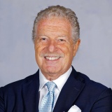 Dr Giovanni Olivi, Italy
Abstract
There has been a volcanic eruption of technologies in Endodontics over the past 20 years, including Laser Activated Irrigation (LAI) from 2007. In recent years, SWEEPS technology was introduced in the market. A dual pulse Er:YAG laser emission produces fluid cavitation in the root canal system as result of the photoacoustic phenomenon generated by ultra-short pulse energy, delivered by a laser tip kept stationary in the access cavity of the tooth. In this way, the use of EDTA and NaOCl, along the correct protocol, improve the cleaning and decontaminating effect of root canals when compared to conventional methods. Differences among the SWEEPS protocol and other erbium laser activated irrigation protocols are underlined, explaining the basic physics of photothermal and photoacoustic effects in dentistry.
Learning Objectives
- Explain the different mechanisms of the various laser techniques used in endodontics.
- Demonstrate the ability of certain techniques to irrigate the entire endodontic system simultaneously.
- Demonstrate the advantage of positioning the laser tip in the access cavity rather than in the canal.
- Present studies comparing the antibacterial efficacy of different disinfection techniques in endodontics.
- Discuss the advantages of LAI versus ultrasonic and SNI techniques for cleaning and disinfecting the root canal system.
Speakers
-
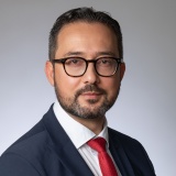 Prof Georgi Tomov, Bulgaria
Abstract
The photodynamic therapy (PDT) has a long history of use and now is a worldwide recognized modality in many fields of medicine like dermatology, oncology and dentistry. A specific kind of PDT is the local antibacterial photodynamic therapy (aPDT) which is considered by some authors as alternative of antibiotic therapy. However, the existing clinical studies are controversial in their conclusions especially regarding its clinical effectiveness. Additionally, there is no consensus on the quantitative measurement of aPDT namely the production of reactive oxygen species (ROS) when different photosensitizers are activated. It also remains unclear how the specific ecological factors in the oral cavity like the local pH, the presence of biofilm matrix etc. could influence the aPDT effectiveness.
Learning Objectives
- The lecture is discussing some important aspects of the local aPDT including evidence based quantitative measurement of ROS production of different photosensitizers.
- The influence of the main oral environmental factors on the clinical effectiveness of aPDT is explained in details.
- The clinical part of the lecture presents the main clinical indications for local aPDT in dentistry.
- Author’s clinical protocol for therapy of peri-mucositis and peri-implantitis is presented and illustrated with relevant clinical cases.
- New potential applications of aPDT are discussed.
Speakers
-
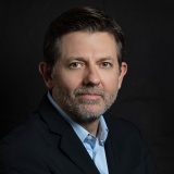 Prof Dimitris Strakas, Greece
-
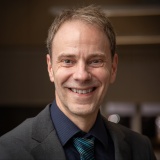 Dr Rene Franzen, Germany
Abstract
Clinical laser dentistry has evolved rapidly across multiple wavelengths, devices, and treatment concepts, but its growth has been accompanied by considerable heterogeneity in terminology, indications, parameter selection, and claims of clinical benefit. Until now, no broad international consensus has attempted to define what is genuinely established across the major domains of clinical laser dentistry and which topics remain unresolved.
This lecture presents the results of the first global Delphi consensus framework in clinical laser dentistry, developed through collaboration between experts from the International Society for Laser Dentistry (ISLD) and the Academy of Laser Dentistry (ALD). Thirty experts with postgraduate education in laser dentistry at MSc level or higher participated in Round 1 and evaluated 120 statements covering laser safety, endodontics, periodontology, operative dentistry and cariology, photobiomodulation, prosthodontics, orthodontics, and oral surgery. Agreement consensus was predefined as ≥70% of participants rating a statement 4 or 5 on a five-point Likert scale.
Round 1 produced consensus for 110 of 120 statements. The 10 unresolved statements were reassessed in a second blinded round by 25 experts, with four additional statements reaching consensus. The final framework therefore achieved agreement on 114 of 120 statements (95%), while six topics remained unresolved. These controversies primarily involved claims of superiority, mandatory treatment sequencing, ceramic debonding, laser surface modification, and specific device-related applications.
Importantly, this initiative was intentionally designed as a foundational consensus rather than an attempt to establish universal treatment protocols or fixed parameter sets. Patient variability, tissue characteristics, clinical indication, wavelength, emission mode, delivery system, and device configuration all limit the validity of one-size-fits-all protocols.
The resulting framework provides a common international basis for education, clinical governance, research reporting, and future wavelength- and indication-specific recommendations. It marks a transition in laser dentistry from technological fragmentation toward structured, evidence-informed global consensus.
Learning Objectives
- Discuss the need for an international consensus framework in clinical laser dentistry and the challenges created by heterogeneous wavelengths, devices, protocols, and clinical indications.
- Explain the Delphi methodology used to develop the first global consensus framework in clinical laser dentistry, including expert selection, consensus thresholds, and the two-round evaluation process.
- Describe the major areas of agreement identified across laser safety, endodontics, periodontology, operative dentistry, photobiomodulation, prosthodontics, orthodontics, and oral surgery.
- Demonstrate an understanding of the distinction between established expert consensus, areas of continuing controversy, and claims that currently lack sufficient support for standardization.
- Explain why universal laser protocols and fixed parameter sets were intentionally not included in this first consensus, considering patient variability, tissue characteristics, wavelength-specific interactions, and differences among laser systems.
- Discuss how the consensus framework can support clinical decision-making, education, research reporting, and the development of future indication- and wavelength-specific recommendations.
- Identify the priority areas for future research and consensus development, including the progression toward evidence-informed therapeutic ranges, safety boundaries, and protocol-level recommendations.
Speakers
-
 Prof Aslihan Usumez, Türkiye
Abstract
Temporomandibular disorders (TMDs) represent one of the most prevalent musculoskeletal conditions affecting the craniofacial region and are a major cause of chronic orofacial pain and functional impairment. Despite the availability of numerous conservative treatment modalities, optimizing pain management while restoring jaw function remains a significant clinical challenge. Photobiomodulation therapy (PBMT) has emerged as a promising non-invasive therapeutic approach owing to its analgesic, anti-inflammatory, and biostimulatory effects. However, considerable variability in treatment protocols has limited the establishment of standardized clinical guidelines.
This keynote lecture will provide a comprehensive overview of the current evidence supporting the use of PBMT in the management of TMDs, with particular emphasis on the optimization of irradiation parameters, including wavelength, power output, energy density, irradiation intensity, treatment frequency, and duration. The biological mechanisms underlying PBMT, including modulation of inflammatory mediators, enhancement of mitochondrial activity, improvement of microcirculation, and stimulation of tissue repair, will be discussed to explain their relationship with clinical outcomes.
Practical recommendations for integrating evidence-based PBMT protocols into routine clinical practice will be presented, together with current limitations of the literature and future research priorities. By translating the growing body of scientific evidence into clinically applicable strategies, this lecture aims to provide clinicians, researchers, and educators with a clear framework for optimizing PBMT protocols, improving patient outcomes, and advancing the management of temporomandibular disorders through precision-based, evidence-driven care.
Learning Objectives
- Describe the pathophysiology of temporomandibular disorders and the rationale for using photobiomodulation therapy.
- Explain how PBMT parameters influence biological responses and therapeutic outcomes.
- Interpret the latest clinical evidence regarding PBMT effectiveness for pain reduction and functional improvement in TMD patients.
- Demonstrate an evidence-based approach to selecting appropriate PBMT treatment parameters for different clinical scenarios.
- Integrate current evidence into clinical decision-making to optimize patient outcomes and improve the quality of TMD management.
Speakers
-
 Dr Miguel Stanley, Portugal
Abstract
Dentistry is undergoing a quiet redefinition — from a discipline of reactive mechanics to one of preventative biology. This keynote explores that shift through the lens of light, and what it means for revision dentistry: the growing clinical reality of treating patients who carry decades of old, outdated dental work — fillings, root canals, crowns, implants — placed under standards and materials we now know were biologically flawed.
That legacy work doesn’t just sit passively in the mouth. Failing margins, low-grade chronic infection at old root canal sites, and materials that were never designed with long-term biocompatibility in mind create a hidden inflammatory burden — one that doesn’t stay contained to the mouth. Drawing on the etymology of “doctor” as teacher rather than technician, Dr. Stanley argues that revision dentistry is not cosmetic touch-up work — it is the removal of a source of systemic biological load that has direct consequences for a patient’s longevity.
Lasers are central to doing this safely. Er:YAG laser disinfection allows compromised sites — old socket infections, failed implant sites, residual bacterial reservoirs left behind by legacy work — to be properly sterilized before any revision procedure, something mechanical instrumentation alone cannot achieve. Low-level light therapy (photobiomodulation) then accelerates regeneration of the tissue that has to rebuild around the revision, reducing the biological cost of correcting decades-old mistakes.
The talk closes by connecting this directly to longevity: every unresolved legacy dental site is a chronic inflammatory input working against a patient’s healthspan. Lasers don’t just make revision dentistry more precise — they make it biologically honest, turning revision from a repeated mechanical fix into a genuine reset of the patient’s oral and systemic health.
“The future of dentistry is not faster. It is wiser.”
Learning Objectives
- Describe the shift from reactive mechanical dentistry to preventative biology, and why revision dentistry is a longevity problem, not a cosmetic one.
- Identify how failing margins, residual infection at old endodontic sites, and non-biocompatible legacy materials create a hidden systemic inflammatory burden.
- Explain the role of Er:YAG laser disinfection in sterilising compromised sockets, failed implant sites, and bacterial reservoirs before revision.
- Apply the principles of photobiomodulation to reduce the biological cost of rebuilding tissue after revision.
- Connect unresolved legacy dental sites to chronic inflammatory load and healthspan.
Speakers
Abstract
The oral-systemic health connection is increasingly important in biological dentistry. Periodontal inflammation and dysbiosis can contribute to a chronic inflammatory burden, and periodontal disease is associated with systemic conditions including cardiovascular disease, diabetes, and adverse pregnancy outcomes. This reinforces the importance of controlling oral infection and inflammation while supporting the body's natural healing response.
Dental lasers provide a biologically oriented approach that may help achieve these goals through reduced tissue trauma, microbial reduction, decreased reliance on medications, and enhanced healing.
The Er laser enables precise hard- and soft-tissue treatment with minimal thermal impact and reduced mechanical trauma. Improved patient comfort, reduced postoperative discomfort and swelling, and potentially less reliance on analgesic medications can support a gentler treatment experience.
Nd and diode lasers offer antimicrobial and soft-tissue applications in periodontal and peri-implant therapy. Their ability to reduce local microbial burden and assist in managing inflamed tissues may support antibiotic stewardship, potentially reducing the need for systemic antibiotics in appropriately selected clinical situations.
Photobiomodulation (PBM) using Nd and diode lasers adds another dimension to biological care by modulating inflammatory responses, supporting cellular activity and tissue repair, and improving postoperative comfort.
By combining infection control, inflammation management, reduced treatment trauma, medication stewardship, and enhanced healing, laser dentistry can complement conventional periodontal and dental therapies within a whole-body philosophy of care.
This presentation explores how Er, Nd, diode lasers, and PBM can move laser dentistry beyond a surgical tool toward a biologically focused therapeutic approach that recognizes the interconnected nature of oral and systemic health.
Learning Objectives
- Discuss the oral-systemic health connection and the relationship between periodontal inflammation and systemic conditions, including cardiovascular disease, diabetes, and adverse pregnancy outcomes.
- Explain how Er:YAG, Nd:YAG, and diode laser wavelengths interact differently with oral tissues and how these characteristics guide their clinical applications.
- Describe how Er:YAG laser applications can reduce mechanical tissue trauma and postoperative discomfort and may help reduce reliance on analgesic medications.
- Discuss the antimicrobial applications of Nd:YAG and diode lasers in periodontal and peri-implant therapy and their potential role in supporting responsible antibiotic stewardship.
- Explain the biological mechanisms and clinical benefits of photobiomodulation (PBM), including modulation of inflammation, support of tissue repair, and postoperative comfort.
- Demonstrate how dental laser applications can be incorporated into a biological dentistry approach focused on microbial control, inflammation management, reduced treatment trauma, and enhanced healing.
Speakers
-
Dr Ilay Maden, UK
Abstract
The oral-systemic health connection is increasingly important in biological dentistry. Periodontal inflammation and dysbiosis can contribute to a chronic inflammatory burden, and periodontal disease is associated with systemic conditions including cardiovascular disease, diabetes, and adverse pregnancy outcomes. This reinforces the importance of controlling oral infection and inflammation while supporting the body's natural healing response.
Dental lasers provide a biologically oriented approach that may help achieve these goals through reduced tissue trauma, microbial reduction, decreased reliance on medications, and enhanced healing.
The Er laser enables precise hard- and soft-tissue treatment with minimal thermal impact and reduced mechanical trauma. Improved patient comfort, reduced postoperative discomfort and swelling, and potentially less reliance on analgesic medications can support a gentler treatment experience.
Nd and diode lasers offer antimicrobial and soft-tissue applications in periodontal and peri-implant therapy. Their ability to reduce local microbial burden and assist in managing inflamed tissues may support antibiotic stewardship, potentially reducing the need for systemic antibiotics in appropriately selected clinical situations.
Photobiomodulation (PBM) using Nd and diode lasers adds another dimension to biological care by modulating inflammatory responses, supporting cellular activity and tissue repair, and improving postoperative comfort.
By combining infection control, inflammation management, reduced treatment trauma, medication stewardship, and enhanced healing, laser dentistry can complement conventional periodontal and dental therapies within a whole-body philosophy of care.
This presentation explores how Er, Nd, diode lasers, and PBM can move laser dentistry beyond a surgical tool toward a biologically focused therapeutic approach that recognizes the interconnected nature of oral and systemic health.
Learning Objectives
- Discuss the oral-systemic health connection and the relationship between periodontal inflammation and systemic conditions, including cardiovascular disease, diabetes, and adverse pregnancy outcomes.
- Explain how Er:YAG, Nd:YAG, and diode laser wavelengths interact differently with oral tissues and how these characteristics guide their clinical applications.
- Describe how Er:YAG laser applications can reduce mechanical tissue trauma and postoperative discomfort and may help reduce reliance on analgesic medications.
- Discuss the antimicrobial applications of Nd:YAG and diode lasers in periodontal and peri-implant therapy and their potential role in supporting responsible antibiotic stewardship.
- Explain the biological mechanisms and clinical benefits of photobiomodulation (PBM), including modulation of inflammation, support of tissue repair, and postoperative comfort.
- Demonstrate how dental laser applications can be incorporated into a biological dentistry approach focused on microbial control, inflammation management, reduced treatment trauma, and enhanced healing.
No programs of this type on this day